
|
|
|
© Dirk Biddle
Particular vasculitis conditions may arise from, or at the very least manifest, different kinds of immune response. The immune response is made up of two cellular systems: the acquired or adaptive,humoral, circulating antibody system (B-cells), and the cell-mediated, innate immune-response system (T-cells).
The cell mediated system acts on antigens appearing on the surface of individual cells. T-cells produce T-cell receptors(TcR) which recognize specific antigens bound to the MHC molecule on the surface of the presenting cell (3).
The humoral immune response is mediated by immunoglobulins. Immunoglobulins are antibodies (antigen recognition proteins) produced by plasma cells (a type of B-cell) that have specific amino acid (protein component) sequences that give each one the ability to adhere to or interact with a specific antigen. It is this antigen-specific property of the antibody that is the essential basis in an immune response. Antibodies may also act as receptors on B-cells. Plasma cell secreted antibody is structurally slightly different than the antibody on the surface of B-cells but the antigen recognition sites are similar.
To achieve the large number of specificities the body needs to protect itself against many different foreign antigens, it must produce millions of B lymphoyctes. It is important to note that, in order to produce such a diversity of antigen binding sites with a separate gene for each possible antigen, the immune system would require many more genes than exist in the genome. Instead, as Susumu Tonegawa showed in 1976, portions of the genome in B lymphocytes can recombine to form all the variation seen in the antibodies and more. Tonegawa won the Nobel Prize in Physiology or Medicine in 1987 for his discovery (4).
Each antibody has two identical heavy polypeptide chains and two identical light chains, shaped to form a Y (see Figure 1). The sections that make up the tips of the Y's arms vary greatly from one antibody to another, creating a pocket uniquely shaped to enfold a specific antigen. This is called the variable (V) region. The stem of the Y serves to link the antibody to other participants in the immune defences (eg; leucocytes). This area is identical in all antibodies of the same class, and is called the constant (C) region (it must be noted that many texts misguidedly refer to the Fc region noted in Figure 1 below as the “constant” region – as if to indicate the “c” in “Fc” stands for “constant”. It does not. The constant region of an antibody is much broader. The “c” actually stands for crystallisable - a reaction of this portion of the antibody under certain laboratory conditions used to cleave and identify the antibody’s components parts, and the whole “Fc” is “crystallisable fragment”). A given antibody matches an antigen much as a key matches a lock. The fit varies: sometimes it is very precise, while at other times it is little better than that of a skeleton key. To some degree, however, the antibody interlocks with the antigen and thereby marks it for destruction. Antigen recognition and binding allows antibodies to perform three important functions: opsonisation, activating complement and neutralizing toxins and toxic organisms.

Immunoglobulins (antibodies) are divided into 5 major classes or isotypes: Immunoglobulin A (IgA - comprising 6% of total antibody), IgD (0.2%), IgE (0.002%), IgG (80%), and IgM (13%).
Interestingly IgA exists as two subclasses: IgA1 (90%) is composed like other antibodies, however in IgA2 (10%) the heavy and light chains are not linked with disulfide but with noncovalent bonds; and IgG exists as four subclasses: IgG1 (66%), IgG2 (23%), IgG3 (7%) and IgG4 (4%) – only IgG4 does not activate complement.
Antibodies function in a variety of ways designed to eliminate the antigen that elicited their production and some antibody functions are independent of the particular class (isotype) of immunoglobulin. These functions reflect the antigen binding capacity of the molecule as defined by the variable and hypervariable (idiotypic) regions. For example, an antibody might bind to a toxin and prevent that toxin from entering host cells where its biological effects would be activated. Similarly, a different antibody might bind to the surface of a virus and prevent that virus from entering its host cell. In contrast, other antibody functions are dependent upon the immunoglobulin isotype. These functions are contained within the constant regions of the molecule. For example, only IgG and IgM antibodies have the ability to interact with and initiate the complement cascade. Likewise, only IgG molecules can bind to the surface of macrophages via Fc receptors to promote and enhance phagocytosis and only IgG can cross the placental barrier, thus providing the foetus with some conferred immunity.
The following briefly summarizes some of the properties of the immunoglobulin isotypes.
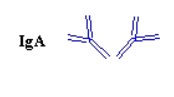
IgA is a type of antibody that protects against infections of the mucous membranes lining the mouth, airways, and digestive tract. This type of antibody is also found in maternal milk, saliva and tears. A small percentage of people do not make IgA antibodies. IgA does not activate complement and opsonises only weakly.
IgA nephropathy (Buerger’s disease) results from deposits of IgA inside the glomeruli of the kidney. These glomeruli (the singular form is glomerulus) normally filter wastes and excess water from the blood and send them to the bladder as urine. The IgA protein prevents this filtering process, leading to blood and protein in the urine and swelling in the hands and feet. This chronic kidney disease (or glomerulonephritis) may progress over a period of 10 to 30 years. If this disorder leads to end-stage renal disease, the patient must go on dialysis or receive a kidney transplant.
Corticosteroids may suppress the production of IgA but can have harmful side effects. In preliminary studies, fish oil supplements containing omega 3 fatty acids also appear to slow the progression of the kidney disease. A new immunosuppressive agent called mycophenolate mofetil (MMF) is also being tested.
A more detailed examination of this vasculitis condition may be found later in this article when we deal directly with the specific vasculitides.
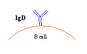
IgD is primarily found on the surface of B-cells and may help regulate B-lymphocyte function (as memory or plasma cells). IgD is co-expressed with IgM on approximately 90% of mature B cells, but its levels in serum are extremely low compared to those of IgM. IgD antibodies are found in small amounts in the tissues that line cavities inside the body. The function of IgD antibodies is not well understood. They appear to play a role in allergic reactions to some substances such as milk, some medications, and some poisons.
The anti-IgD antibody may also help identify autoimmune diseases such as glomerular disease in Systemic Lupus Erythematosis (SLE) causing acute or chronic glomerulonephritis.
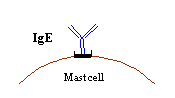
The protective role for IgE is not clear but IgE antibodies are found in the lungs, skin, and mucous membranes. They cause the body to react against foreign substances such as pollen, fungus spores, and animal dander. IgE may also play a role in the defence against parasites such as worms. IgE antibody levels are often high in people with allergies. IgA attaches to cell membranes causing the release of histamine and other substances responsible for the local inflammation characteristic of an allergy. IgE does not activate complement and of all antibody isotopes only IgE is heat-labile.
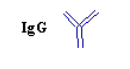 IgG antibodies are found in all body fluids. They are the smallest but most abundant of the antibodies. IgG coats bacteria, helping other cells (eg; eosinophils) to seek and destroy them. IgG antibodies are considered the most important antibodies for fighting bacterial and viral infections. IgG antibodies are the only type of antibody that can cross the placenta. Therefore, the IgG antibodies of a pregnant woman can also help protect her foetus.
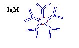
IgM antibodies are the largest type of antibody. They are found in blood and lymph fluid and are the first type of antibody produced in response to an infection. They also cause other immune system cells to produce compounds that can destroy invading cells. IgM is particularly efficient in activating the complement cascade. IgM has also been termed a "natural antibody" as it is found in the serum without any evidence of prior contact with antigen.
The association between various immunoglobulins and vasculitis conditions is the subject of a great deal of research but remains not well understood. It is clear however that some vasculitis conditions are strongly associated. For example IgA with Henoch-Schönlein purpura, Berger’s disease and Cutaneous leukocytoclastic angiitis. IgA deposition is also often noted in Glomerulonephritis (5, 6), sometimes IgM, but rarely IgG. Further, IgG is dominant in Systemic Lupus Erythematosus (5) and IgG and IgM are strongly associated with cryoglobulinemia (8, 9).
Some vasculitis conditions are not associated at all (or very weakly associated): for example, Microscopic polyangiitis, Wegener granulomatosis, Churg-Strauss syndrome, and renal-limited pauci-immune crescentic glomerulonephritis - for which few if any immune deposits can be identified in target tissues (7).
However it is also true that many (if not all) vasculitis conditions are associated (at least) in some small way with immune deposition. Thus IgA deposition may also be found in most ANCA positive vasculitis (5) including Berger’s disease, CNS vasculitis (10), Polyarteritis Nodosa, Wegener’s granulomatosis, Sjögren's syndrome, and Systemic Lupus Erythematosus. IgD is rare in vasculitis conditions but has been associated with Henoch-Schönlein purpura (11) and Urticaria (12) in association with hyperimmunoglobulinemia D Syndrome. IgE may be involved in cutaneous leukocytoclastic vasculitis (13) and has been noted in Churg-Strauss syndrome (8) and Urticaria (14), and so on.
Thus the picture of association is extremely complex and more on these links will be forthcoming when we come to describe the particular vasculitis conditions in more detail in later sections.
----------------------
| back |
Humoral:Of, relating to, proceeding from or involving a bodily humour - now often used of endocrine factors as opposed to neural or somatic. (OMD)
1: of, relating to, proceeding from, or involving a bodily humor (such as a hormone)
humoral control of sugar metabolism2: relating to or being the part of immunity or the immune response that involves antibodies secreted by B-cells and circulating in bodily fluids. (M+),
Cell-Mediated Immunity: Immune response that involves effector T-lymphocytes and not the production of humoral antibody.
Responsible for allograft rejection, delayed hypersensitivity and in defence against viral infection and intracellular protozoan parasites. (OMD)
Relating to or being the part of immunity or the immune response that is mediated primarily by T cells and especially cytotoxic T cells rather than by antibodies secreted by B cells. (M+),
T-Cell Receptors: Any of several lymphocytes (as in helper T-cell) that differentiate in the thymus, possess highly specific cell-surface antigen receptors, and include some that control the initiation or suppression of cell-mediated and humoral immunity (such as by the regulation of T- and B-cell maturation and proliferation) and others that lyse antigen-bearing cells - called also T-lymphocyte. (M+),
Antigen: A (usually) protein or carbohydrate substance (as a toxin or enzyme) capable of stimulating an immune response. (M+),
Immunoglobulin: (Ig) see Antibody. Types include: IgA, IgG, IgB, IgD, IgE, and IgM. Most immunoglobulins are IgG (they migrate in the gamma region during electrophoresis),
Antibody: Any of a large number of proteins of high molecular weight that are produced normally by specialized B-cells after stimulation by an antigen and act specifically against the antigen in an immune response, that are produced abnormally by some cancer cells, and that typically consist of four subunits including two heavy chains and two light chains - called also immunoglobulin. (M+),
Amino Acid: An amphoteric organic acid containing the amino group NH2 (M+) - and a carboxyl group (COOH). Twenty alpha-amino acids are the subunits which are polymerised to form proteins. (OMD),
Antigen: A (usually) protein or carbohydrate substance (as a toxin or enzyme) capable of stimulating an immune response. (M+),
Polypeptide: A molecular chain of amino acids. (M+)",
Opsonisation: A process through which a cell or microbe is treated with opsonin to make it more vulnerable to being engulfed by a phagocyte. (OMD),
----------------------
1. Kossard, S. (2000) Defining lymphocytic vasculitis. Australasion journal of Dermatology. 41(3), 149-155.
2. Smoller, B., McNutt, N., & Contreras, F. (1990) The natural history of vasculitis. What the histology tells us about pathogenesis. Archives of Dermatology. 126(1).
3. (http://wsrv.clas.virginia.edu/~rjh9u/imresp.html)
4. (http://en.wikipedia.org/wiki/Immunoglobulins)
50. (http://www.uninet.edu/cin2003/conf/ferrario/ferrario.html)
6. (http://www.medschool.lsuhsc.edu/pathology/pathist/RENAL/GLOMERULOPATHIES/1MSGN/msgn_references.htm)
7. http://www.jci.org/cgi/content/full/110/7/919
J. Clin. Invest. 110:919-921 (2002). doi:10.1172/JCI200216699.
Copyright ©2002 by the American Society for Clinical Investigation
8. (http://www.dokkyomed.ac.jp/dep-k/cli-path/a-super/vasculitis/super-v-04.html)
9. (http://www.orpha.net/data/patho/GB/uk-cryoglobul.html)
10. Petrasek, J., Panitch, H., Wofsy, D., et al. (1980) IgA-Associated Granulomatous Angiitis (GA) of Brain: Successful Therapy With Cyclophosphamide. Neurology, 30, 442.
11. (http://www.thedoctorsdoctor.com/diseases/hyper_igd_syndrome.htm)
12. (http://www.aaaai.org/aadmc/ate/fever.html)
13. (http://pmj.bmjjournals.com/cgi/content/full/78/916/114)
14. http://www.news-source.org/ACAAI/Archive/FellowsInTraining12-23-03.htm